
For example, in a 2D abdominal ultrasound examination, it’s important to visualize the parenchyma of the organ. If the structure is homogeneous or heterogeneous with surface nodularity, the echogenicity, boundary borders between different organs, tissue differences and focal changes. To differentiate focal changes is fundamental. A simple cyst is typically round or oval, anechoic, and has smooth, thin walls.
What defines image quality in 2D and 3D ultrasound?
I’m a sonographer with a clinical background from ultrasound. I’ve learned how important it is to really know how to use the ultrasound equipment and applications, to provide good care for the patient. Working with diagnostic ultrasound for many years also made me understand the significance of image quality.
In three-dimensional ultrasound, a clear, representative image of the fetal anatomy can be achieved. The use of 3D imaging has already been established for prenatal ultrasound in general and for improving the diagnostic accuracy in fetal neurosonography and echocardiography.

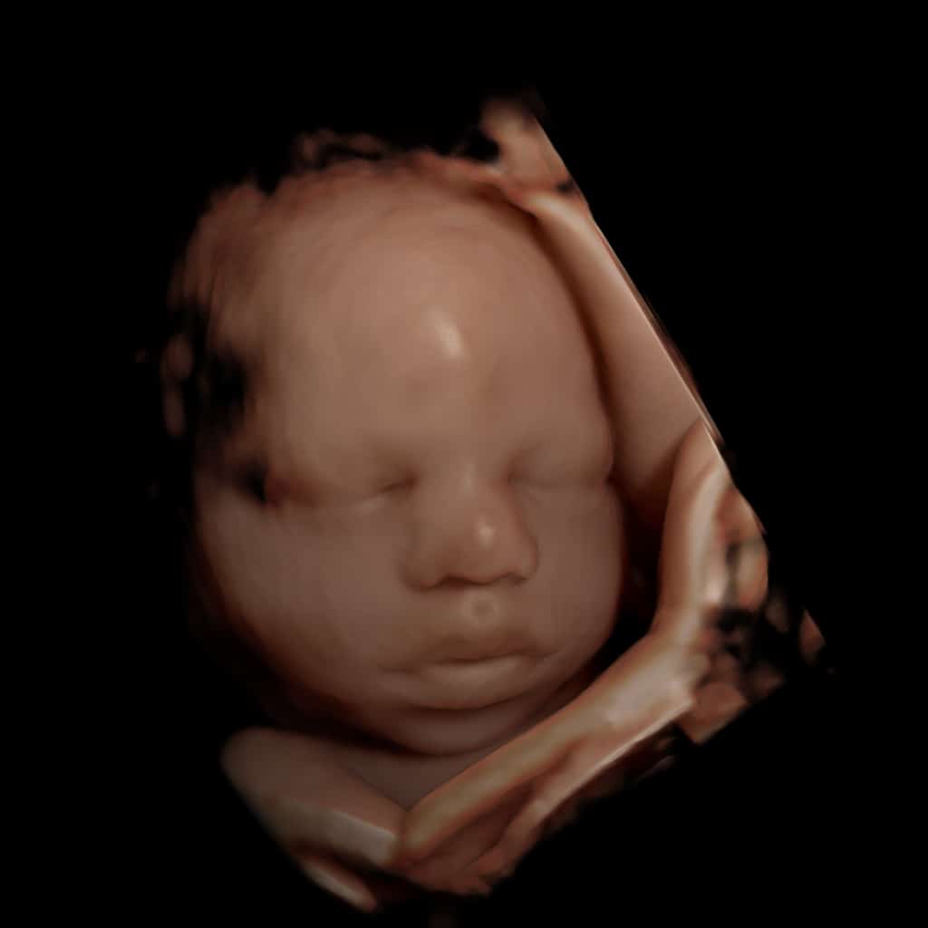
Processed with Rivent 3D
In prenatal diagnosis the face is important to evaluate, and here, 3D ultrasound gives higher accuracy in assessing the fetal face and profile, including the nose, chin, and forehead as well as a coronary en-face view of the mouth. More precise measurements in 3D multiplanar volumes facilitate diagnosis of micrognathia, syndromes, and skeletal dysplasia.
How much or how little ultrasound experience the examiner has, has a significant impact on the detection rate of various diagnoses. The level of certainty depends on the experience and expertise of the examiner. However, in-depth knowledge of how to operate the ultrasound equipment is essential for optimal image adjustment. From the standard setting and the presets on the ultrasound machine, it’s possible to adjust a few parameters. For example, frequency, depth in the image, gain, focus and dynamic range. But changing these parameters isn’t always enough to achieve a good image. Some patients are more difficult to examine than others, so the examiner must also know how to position the patient, choose the right transducer, etc.
Of course, what defines a good ultrasound image is subjective.
But from a medical point of view, the examiner’s goal should always be to achieve the best possible image quality.
Learn more
Ut enim ad minim veniam, quis nostrud exercitation ullamco laboris nisi ut aliquip ex ea commodo consequat.
What does research mean at ContextVision?
Lorem ipsum dolor sit amet, consectetur adipiscing elit, sed do eiusmod tempor incididunt ut labore et dolore magna aliqua. Ut enim ad minim veniam, quis nostrud exercitation ullamco laboris nisi ut aliquip ex ea commodo consequat.
Read more →
People make AI intelligent – that’s our philosophy
Consectetur adipiscing elit, sed do eiusmod tempor incididunt ut labore et dolore magna aliqua. Ut enim ad minim veniam, quis nostrud exercitation ullamco laboris nisi ut aliquip ex ea commodo consequat.